
OJVRTM
Online Journal of Veterinary Research©
Volume 7 : 26-32,
2003.
Arun KHS, Manjuna HS1, Reddy KS, Reddy BA.
Department of Pharmacology and
Toxicology,
College of Veterinary Science, ANGRAU, Rajendranagar, Hyderabad, India.
@ Dept of LPM. 1Biochemist,
Biocon India Ltd., Bangalore, India.
Arun KHS, Manjuna HS, Reddy KS, Reddy BA. Sub-acute oral toxicity of Salinomycin in broiler chicks Online Journal of Veterinary Research 7 : 26-32, 2003. The effects of 120 ppm salinomycin in feed (sub-acute oral toxicity) on clinical signs, tissues and serum biochemistry of broiler chicks was evaluated. Symptoms ranged from in-coordination, lethargy, leg weakness, diarrhea, reduced feed intake and weight loss. Sternal recumbency with neck, wings and hind limbs outstretched were characteristic and prominent by the 4th week of salinomycin exposure. Extensive pathology occurred in skeletal muscle, heart, lungs, kidney, liver, intestines and bursa. Serum alanine aminotransferase, aspartate aminotransferase, alkaline phosphatase and lactase dehydogenase, urea, creatinine and cholesterol increased whereas total protein, albumin and globulin decreased. There were no significant changes in values taken from control birds or those fed 60 ppm salinomycin.
KEY-WORDS: Salinomycin, biochemical parameters, and toxicity.
Polyether ionophores in feed prevent losses due to coccidiosis in broiler chicks raised on deep litter (Folz et al. 1988; Elwinger et al. 1998). Unfortunately, polyether ionophore's narrow margin of safety and uneven distribution in feed is a major disadvantage (Dowling 1992; Rizvi & Anjum 1999). Salinomycin is a polyether ionophore drug extensively used to control coccidiosis because of its broad spectrum of activity and slow development of resistant strains (Mc Dougld & Roberson, 1988). Salinomycin is a monovalent polyether fermentation product produced by a strain of Streptomyces albus (Kinashi et al., 1973). It is available as a 12 % premix, for use in broiler feed and is recommended @ 44-66 ppm in feed (Folz et al., 1988). Though salinomycin is least toxic among all the ionophores presently available, certain untoward effects such as reduced feed intake and depressed growth in chicks (fed on recommended levels of salinomycin) have been reported in the absence of coccidiosis.
The toxic effects of
salinomycin
are obvious at high doses due to improper mixing, or sedimentation and
segregation in feed during storage causing high levels in the lower
part
of the grain (Rizvi & Anjum,
1999).
Braunius (1985) and Dowling,
(1992) showed that 20-50% over-dosage induced incoordination, leg
weakness,
diarrhea, reduced feed intake and weight depression. Diagnosis of
ionophore
toxicity is uncertain since clinical signs and lesions are not
pathognomic
and can be reversible. Confirmatory diagnosis requires differential
diagnoses
and laboratory assays of tissue and feed. The present study describes
the
effects of salinomycin sub-acute toxicity on serum biochemical
parameters
and tissues in chick broilers
Animal ethics:The trial was approved by the Institutional Animal ethics Committee, ANGRAU, Hyderabad complied with Halenski's declaration on handling of experimental animals.
Chicks:Thirty male on day old broiler chicks (Venkateswara Hatcheries, Hyderabad) were randomly divided into 3 groups 10 each. The birds were vaccinated against New Castle Disease (NDV; day 7 & 21), Infectious Bronchitis (IB, day 1) and Infectious Bursal Disease (IBD, day 14 & 23) as per the schedule recommended by the Venkateswara Hatcheries, Hyderabad, India. All the birds were housed under a cage system in a well ventilated broiler shed with ad-lib feed and water access. Chicks of group I served as control without coccidostat in the feed, groups II and III birds were fed with silinomycin @ 60 (S60) and 120 ppm (S120) respectively in feed (the feed was prepared in the institute farm feed milling unit as per the composition given in table below).
Feed formula table:
| Ingredients | Kgs |
| Maize (Yellow) | 63.16 |
| Soya | 33.28 |
| Di-calcium phosphate | 1.35 |
| Lime Stone | 1.4 |
| DL methionine | 0.15 |
| AB2D3K | 0.01 |
| BE | 0.01 |
| B12 | 0.01 |
| Choline Chloride | 0.05 |
| Probiolac | 0.01 |
| Trace Minerals | 0.10 |
| Coccidostat (Salinomycin) | 60 or120 ppm |
Body weights and Feed Conversion efficiency (FCR):The growth pattern was studied by recording the weekly body weights (0-6 weeks) of all the birds in each group and the average body weight was calculated for each group. FCR was calculated using the following formula:FCR = Total feed consumed / total body weight gained.
Biochemical studies: Blood samples were drawn from wing vein once every fortnight using a 24-gauge needle attached to 2-ml syringe for 6 weeks. The blood collected was immediately transferred to a glass test tube along its sides and was kept in slanting position for the serum separation (a small quantity of blood was immediately used for the hematological studies). Serum separated was used for the assay of alanine aminotransferase {ALT} (King, 1962), aspertate aminotransferase {AST} (King, 1962), alkaline phosphatase {ALP} (Kind & King, 1954), total protein (Doumas, 1978), albumin (Doumas, 1978), globulin (Doumas, 1978), urea (Natelson, 1957), creatinine (Bartels et al., 1972), cholesterol (Charles et al., 1974), triglycerides (Bucolo & David, 1973) and lactate dehydrogenase {LDH} (Henry & Winkelman, 1974) using standard kits procured from Bhat Biotech (I) Ltd., Bangalore India.
Hematological studies: Hemoglobin (Hb), packed cell volume (PCV), total erythrocyte count (TEC), total leukocyte count (TLC) and platelet count were estimated using automated Animal Blood Counter (Sos-Icsaquoits).
Histopathological studies: A detailed post-mortem (toxicopathological) examination of birds sacrificed in each group was conducted at the end of sixth week. The gross pathological changes, if any were noted. Tissue samples from liver, heart, kidney, skeletal muscles, intestine, bursa and lungs were collected for histopathological studies by Hematoxyline and Eosin (H&E) staining (Singh & Sulochana, 1997).
Organ index: Organ index was estimated for the liver, gizzard, pro-ventriculus, kidneys, heart, lungs and spleen, using the following formula: Organ Index = Organ weight (g) / Live body weight (g)
Materials: Salinomycin (Coxistac 12 % premix) was provided by Pfizer Ltd.; Mumbai, NDV, IB and IBD vaccine were procured from Venkateswara Hatcheries, Hyderabad, H & E stain was procured from MS Qualigens Pvt. Ltd., Hyderabad. Feed ingredients were procured form the authenticated suppliers to the poultry farm, ANGRAU, Hyderabad. All the kits for biochemical studies were procured from Bhat- Biotech India Pvt., Ltd. Bangalore.
Statistical
analysis: All the values are expressed as Mean ±
SD. The data was analyzed by two-way analysis of variance (ANOVA),
using
Jandel SigmaStat 2, statistical software followed by multiple groups
comparison
with Tukey`s test or Bonferroni`s test. The values were considered
significant
at P<0.05.
RESULTS
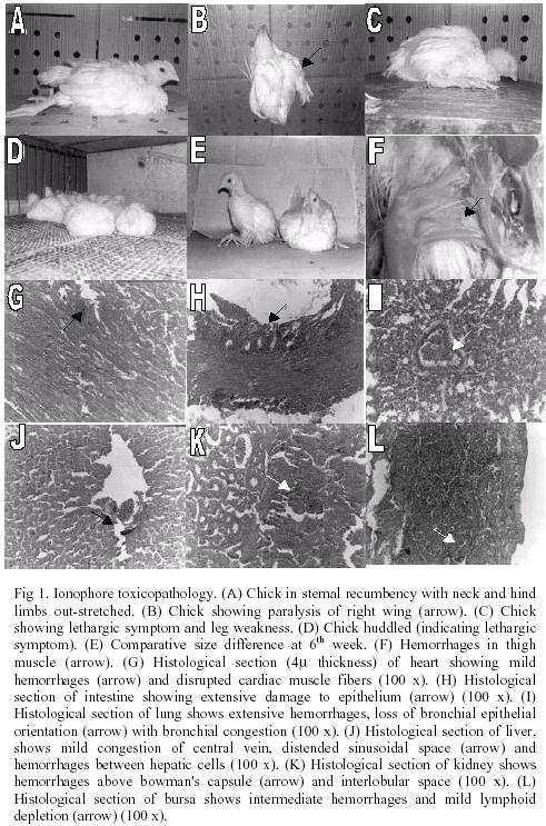
Salinomycin treated (120-ppm) chicks showed symptoms of toxicity, which ranged from in-coordination, lethargy, leg weakness, diarrhea, reduced feed intake and weight loss. Birds in sternal recumbency with neck, wings and hind limbs outstretched were very characteristic and prominent after 4th week of salinomycin exposure. However, the S60 and control group birds did not show these symptoms throughout the study.
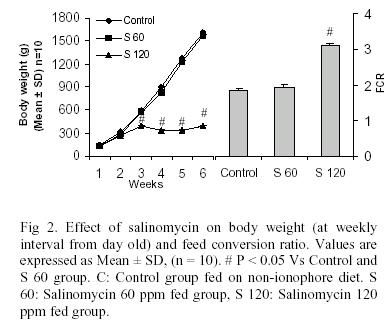
The weekly average body weight
(g)
and feed conversion ratio (FCR) of birds in all the groups are
expressed
as mean ± SD in table 1 (fig 1).
The average body weights of control and S60 group was significantly (p < 0.05) higher than S120 group, however the difference in body weights between control and S60 group was non-significant. This difference in body weights was evident as early as first week onwards and continued until termination of the study (6th week). The FCR was significantly higher (p < 0.05) in S120 group as compared to control and S60 group. While the difference in FCR between control and S60 group was non-significant (table 1, fig 1).
The hematological profiles (Hb, TEC, TLC, platelet count, PCV, MCV, MCH and MCHC) didn't show any significant (p > 0.05) difference between the various groups at 2nd week but was significantly lower in S120 group from 4th week onward as compared to control and S60 group. However, the differential leukocyte count (DLC) didn't show any significant change among the various groups (table 2).
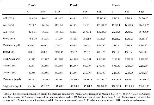
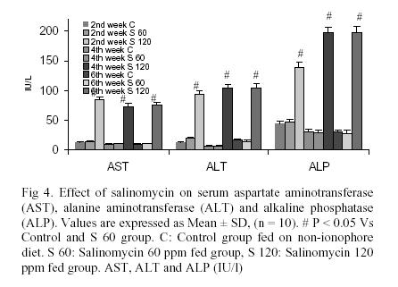
Serum AST, ALT, ALP, (fig 2) LDH (fig 3) activity was significantly (p<0.05) elevated as weeks progressed in salinomycin (120 ppm) treated chicks (Table 3), while the levels were well with in the normal range in to control and S60 group.
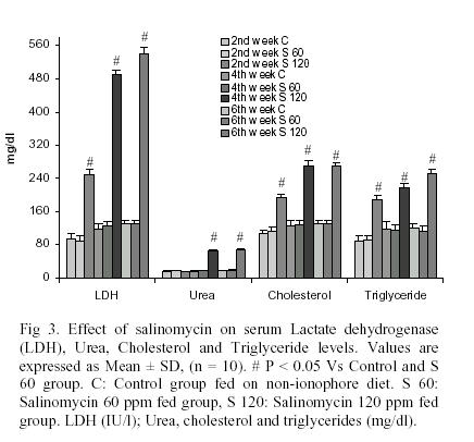
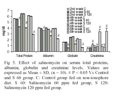
Total protein, albumin, globulin (fig 4) and HDL levels were significantly lower in S120 as compared to control and S60 group. While serum creatinine, cholesterol, triglycerides and LDL levels were significantly (p<0.05) higher in salinomycin (120 ppm) treated chicks (table 3).
Serum urea levels didn't show any change between the various groups at 2nd week, however from 4th week onwards serum urea levels were significantly (p<0.05) higher in salinomycin (120 ppm) treated chicks (group III) as compare to control and S60 group (table 3, fig 2).
Postmortem findings:
The postmortem findings were
typical
of any generalized toxicity with varying pathological signs in various
organs. Hemorrhages in the skeletal muscle (thigh muscle) and heart
(with
extensive dilatation of coronary vessels) were observed in birds of
S120
group. Apart from these, petichiae in liver, kidneys and lungs were
also
noticed. The birds from control and S60 group didn't show any of the
above
pathological changes.
Organo somatic index:
Organ index of heart and liver
were
significantly (p < 0.05) higher in S120 group as compared to control
and S60. While the organ index of kidneys, lungs, proventriculus,
gizzard,
intestines and spleen differed non-significantly (p > 0.05).
Histopathological findings:
The organ wise
histopathological
changes observed in S120 group was as follows:
Heart: Mild hemorrhages with
extensive
disruption of cardiac muscle fiber was observed.
Intestines: Extensive damage to
epithelium with slight to moderate damage of mucosa, a submucosa and
muscularis
layer was seen.
Lungs: Extensive hemorrhages and
loss of bronchial epithelial orientation with bronchial congestion was
noticed.
Liver: Mild congestion of central
vein, distended sinusoidal space and hemorrhages between hepatic cells
was observed.
Kidney: Hemorrhages above bowman's
capsule and focal hemorrhages in interlobular space was observed.
Bursa: Interfollicular hemorrhages
and mild lymphoid depletion in few a follicles was seen.
DISCUSSION
Though polyether ionophore use in broiler feeds is indispensable (Folz et al., 1988; Elwinger et al., 1998), their narrow safety margin is a cause for concern taking into accounts the relative beneficial Vs risk ratio (Oehme & Pickrell 1999). This ratio shifts to its right when the farm management practices are overlooked or compromised giving rise ionophore toxic episodes. A sudden outbreak of salinomycin toxicity in male breeder turkeys (21.7% mortality) with no significant gross lesions and degeneration and necrosis of skeletal muscle was reported due to an error at the feed milling unit (Andreasen & Schleifer, 1995) such errors are as well reported in many broiler farms in and around Hyderabad, India resulting in huge economic loss this prompted us to take up the present study.
Since ionophore toxicity is diffused (not involving any particular organ system) and hence may involve many biochemical changes, we designed this study to evaluate the toxic effect of one such ionophore, salinomycin on serum biochemical parameters as understanding of the biochemical changes would greatly help in adopting suitable remedial measures to avoid such toxic episodes and thus prevent the associated economic loss in broiler farm. The higher salinomycin dose was selected based on a preliminary study employing various doses of salinomycin in feed considering the literature (Matabudul et al. 2002) and as well from field surveillance data on the levels of salinomycin in various feed samples obtained from different broiler farms in and around Hyderabad, India (data not shown).
Salinomycin treated (120-ppm) chicks showed symptoms of toxicity, which ranged from in-coordination, lethargy, leg weakness, diarrhea, reduced feed intake and weight loss. Birds in sternal recumbency with neck, wings and hind limbs outstretched were very characteristic and prominent after 4th week of salinomycin exposure. Similar symptoms of slinomycin toxicity were reported in broiler chicks (Dowling, 1992). Salinomycin administered at 0.25, 1 or 4 times above their recommended dose levels didn't significantly induced tibial dyschondroplasia (TD) in growing boilers (Peters et al., 2002) hence the paralysis observed in salinomycin toxicity could be due its direct effect on skeletal muscles. Control and S60 group chicks didn’t exhibit any of these symptoms. However, a feed-related problem characterized clinically by anorexia, diarrhea, dyspnea, ataxia, depression, recumbency and death, and pathologically focal degenerative cardiomyopathy, skeletal muscle necrosis, and congestive heart failure has been reported to be a presumptive diagnosis of ionophore toxicity (Hoop, 1998) and is in concurrence with our study. Birds fed maduramicin are reported to drink more water than the nonmedicated birds (P<0.05), and polydipsia was reported to be much more in birds fed salinomycin (P<0.05) (Radu et al., 1987) however, we didn't observe any such changes in water in take in S60 or S120 group.
Change in body weight and feed conversion ratio (FCR) are important markers of broiler health and economy. In the present study, that the average body weights of salinomycin (120 ppm) treated group was significantly (p < 0.05) lower than C and S 60 groups. Most of the growth depression with salinomycin diets can be attributed to reduced feed consumption due its anorexic property (Lehel et al., 1995a; Migaki & Babcock, 1979) and is in consistence with the previous reports (Keshavarz & McDougald, 1982). Few reports have shown weight gains at 50 and 60 ppm of salinomycin and 80 and 100 ppm of salinomycin, which was statistically comparable (equivalent) to or better than controls (Migaki & Babcock, 1979). However, the weight gain at 80 ppm of salinomycin was slightly below the control group but at 100 and 160 ppm salinomycin was reported to depress weight gain while FCR for all treatments except 160 ppm salinomycin were comparable (Migaki & Babcock, 1979). Contradictory to these reports certain studies indicate no effect of salinomycin at higher doses on final body weight, and rather have reported an improvement in FCR (Radu et al., 1987).
Prolonged use of salinomycin even at recommended dose as prophylactic measure against coccidosis has been reported to result in growth suppression (Rizvi & Anjum, 1999), however certain reports indicate no adverse effects of salinomycin (40-80 ppm) on body weight in broilers (Pearson et al., 1990). In the present study salinomycin at 120 ppm and not 60 ppm was found to be marked anorexogen and hence resulted in a poor feed conversion ratio. Our results are in accordance with the previous reports in broiler birds (Dowling, 1992; Rizvi & Anjum, 1999). Though we found salinomycin 60 ppm treated birds weighing slightly less, there wasn't any change in FCR among control and S60 group indicating weight gain is indeed affected by salinomycin even at the recommended dose. One of the factors responsible for variable reports on growth depression induced by ionophores could be the incompatibility of micro and macro feed ingredients with the ionophore (Prohaszka et al., 1987) and/or the variability in the strain of the broiler breed used for the study. Saprophytic bacteria during excessive multiplication in poultry feed release metabolites, which have been reported to enhance the toxicity of salinomycin (Prohaszka et al., 1987). Hence we emphasis that it is extremely important to ensure the quality and standard of feed regularly (especially when using the stored feed) to avoid the incidences of ionophore toxicities (and the resulting losses) in the broiler farm.
To establish the nature of biochemical change and weather any the biochemical markers would be ideal in diagnosing ionophore toxicity, we measured various serum biochemical makers once every fortnight until 6 weeks of age. Since the maximum age of broiler marketing in our farm is not beyond 6 weeks, we didn't proceed beyond 6 weeks in the present study. Among the biochemical parameters, tested serum aspartate aminotransferase (AST) activity was significantly elevated as weeks progressed in salinomycin (120 ppm) treated chicks (Table 1; Fig 2) where as it remained normal in the other two groups. Highest AST activity in chicks occurs in heart muscle followed by liver and skeletal muscle and an elevation in its activity in serum has also been associated with hepatocellular damage (Campbell & Coles, 1980), whereas a moderate increase in its activity is associated with other soft tissue injuries. Increased serum AST activity was most prominent at 6th week (Fig 4) and was well correlated with the histopathological findings. Heart, skeletal muscle and liver have low alanine aminotransferase (ALT) activity in chicks (Campbell & Coles, 1980) and it is not generally considered as ideal marker in chicks. However, we observed a significant (p<0.05) elevation in ALT activity in S120 group (Table 1) and this could be due to hepatic damage (Kaneko et al., 1997) caused by toxic dose of salinomycin.
A significant (p<0.05) elevation in serum lactate dehydrogenase (LDH) activity was observed in S120 group (Table 1; Fig 2). Highest LDH activity in chicks has been observed in skeletal muscle followed by that in heart muscle, liver and lungs, however these are different sub types of LDH depending on its location. Elevated serum LDH activity is indicative of hepatic damage though not specific as the damage to heart, lungs, kidneys, intestines or skeletal muscle may also elevate serum LDH activity (Kaneko et al., 1997). Since we have not estimated the sub type of LDH being elevated, we cannot conclude the observation with any particular organ damage. Serum ALP activity was significantly (p<0.05) increased in S120 group (Table 1), which might be due to intestinal disturbance and in-appetence caused by toxic levels of salinomycin (Campbell & Coles, 1980). Serum cholesterol and triglyceride levels were significantly elevated in S120 group (Table 1), this could probably be due to anorexia (Lehel et al., 1995a,b) and/or liver damage (Kaneko et al., 1997) caused by toxic levels of salinomycin.
Hypo-proteinemia has been associated with chronic renal or hepatic damage and/or malnutrition or malabsorption syndrome (Kaneko et al., 1997). A significant decline in serum total protein and albumin levels observed in S120 group (Table 1) could be due to anorexic property of salinomycin (Lehel et al., 1995a,b) thus resulting in malnutrition. We observed the birds being lethargic and away from the feeders for most of the time. Hypoproteinemia observed in S120 group may also be due to toxic effects of salinomycin on kidney, liver and intestines resulting in malabsorption of nutrients. A significant (p<0.05) decrease in serum globulin levels in S120 group (Table 1) may be a consequence of hypoproteinemia or an indicative of immmunosuppression caused by toxic dose of salinomycin. We are presently investigating the effect of polyether ionophores on immune responses in broilers.
Serum urea nitrogen and
creatinine
levels were significantly (P<0.05) elevated in S120 group (Table 1)
which might be due to kidney damage affecting the filtration mechanism
(Kaneko et al., 1997).
Though the changes in the
biochemical
markers is ubiquitous and cannot be specifically associated with any
particular
organ damage, to avoid this confusion we compared the biochemical
changes
with the postmortem and histopathological findings. The postmortem
changes
correlated well with various biochemical changes and the
histopathological
findings further support the organ damage. From the literature
available,
we couldn't find any study reporting detailed histopathological changes
in salinomycin toxicity. Hence, ours is the first such study reporting
gross histopathological changes in various organs in salinomycin
toxicity.
In addition, reports on changes in biochemical parameters in
salinomycin
toxicity are greatly lacking here for the first time we report changes
in various biochemical markers due to salinomycin toxicity and further
we correlate these biochemical markers with postmortem and
histopathological
changes. Presently we are further investigating the changes in
biochemical
markers in the specific organs to have better correlation with
salinomycin
toxicity.
Cytochrome P-450 and Liver malondialdehyde concentration (an indicator of oxidative stress) was reported to be elevated in salinomycin-intoxicated birds. Further a decrease in hepatic glutathione concentration and glutathione peroxidase activity and increase in hepatic catalase activity was reported. In an another study manifestation of the effect exerted by salinomycin and salinomycin-tiamulin on lipid-peroxidative processes was reported to coincided with the onset of clinical signs and preceded the increase of hepatic cytochrome P-450 concentration (Mezes et al., 1992). Salinomycin was reported to be fetotoxic when inoculated into embryonated chicken eggs. The dead fetuses in this study were haemorrhagic, dwarfish and friable while the surviving fetuses showed reduced body weight, insignificant decrease in leg and wing lengths and many developmental abnormalities (Atef et al., 1989). Calcium ionophore have been shown to be neurotoxic effects, which is due to the rise in cytosolic calcium (extracellular calcium influx) (Veinbergs et al., 2002). These report point towards the involvement of free radicals in salinomycin toxicity, this concept of free radicals involvement in salinomycin toxicity could be rational looking in to the diffuse damage seen involving all the organs studied, further the enhanced cytosolic calcium may be a key factor in the free radical generation, we are presently exploring in this area.
From the results of present
study
it can be concluded that salinomycin at 120 ppm induces damage to
skeletal
muscle, kidneys, liver, heart, intestines, lungs and bursa with
corresponding
changes in the biochemical parameters. Hence, the ionophore induced
damage
is ubiquitous. However, the biochemical changes are not specific and
sensitive
indicators of salinomycin toxicity as the damage is not limited to a
particular
organ system, hence diagnosis should be based on postmortem finding
correlated
with feed analysis for ionophore levels. All the parameters studied
remained
normal in control and salinomycin (60 ppm) supplemented group.
Presently,
there is no antidote or treatment for toxicoses induced by the
ionophores.
Judicious use, avoidance of overdosing, and adherence to species
recommendation
will help prevent the occurrence of adverse effects associated with
this
class of compounds (Novilla, 1992). Hence, it is advised to take
adequate
care while mixing salinomycin in feed to ensure its appropriate levels
and avoid using feeds stored for long time without its proper mixing
before
use.
We are thankful to Acharya
N.G.Ranga
Agricultural University for providing necessary facilities to carry out
this study. Dr. Arun Kumar HS was awarded Col Robert Gabriel
Scholarship
from the Ministry of Defense during his MVSc program.
Andreasen,
J.R., Jr. & Schleifer, J.H. (1995). Salinomycin toxicosis in male
breeder
turkeys. Avian Dis, 39, 638-42.
Atef, M.,
Shalaby, A.A., Khafagy, A. & Abo-Norage, M.A. (1989). Fetotoxicity
of some anticoccidial drugs in chickens. Dtsch Tierarztl Wochenschr,
96,
296-8.
Bartels,
H., Boehmer, M. & Heierli, C. (1972). Serum creatinine
determination
without deproteinization. Clinical Chemistry Acta, 37, 193-197.
Braunius,
W. (1985). Ionophore anticoccidial drugs in coccidiosis control. World
Poultry Science Journal, 136, 765-768.
Bucolo,
G. & David, M. (1973). Quantitative determination of serum
triglycerides
by the use of enzymes. Clinical Chemistry, 19, 476-482.
Campbell,
T.W. & Coles, E.H. (1980). Avian clinical pathology:. In Veterinary
clinical pathology. ed. Coles, E. pp. 279-297. Philadelphia: WB
Saunders
Company.
Charles,
C.A., Lvey, S.P., Cieely, S.G., Richmond, W. & Paul, F.C. (1974).
Enzymatic
determination of total serum cholesterol. Clinical Chemistery, 20,
470-475.
Doumas,
B.J. (1978). Biuret and BCG dye method. Clinical Chemistry, 21, 1159.
Dowling,
L. (1992). Ionophore toxicity in chickens: A review of pathology and
diagnosis.
Avian Pathology, 21,, 355-368.
Elwinger,
K., Berndtson, E., Engstrom, B., Fossum, O. & Waldenstedt, L.
(1998).
Effect of antibiotic growth promoters and anticoccidials on growth of
Clostridium
perfringens in the caeca and on performance of broiler chickens. Acta
Vet
Scand, 39, 433-41.
Folz,
S.D., Lee, B.L., Nowakowski, L.H. & Conder, G.A. (1988).
Anticoccidial
evaluation of halofuginone, lasalocid, maduramicin, monensin and
salinomycin.
Vet Parasitol, 28, 1-9.
Henry,
R.J. & Winkelman, J.W. (1974). Clinical Chemistry- Principles and
techniques.
New York ,: Harper and Row.
Hoop,
R.K. (1998). Salinomycin toxicity in layer breeders. Vet Rec, 142, 550.
Kaneko,
J.J., Harrey, J.W. & Micher, L.B. (1997). Clinical biochemistry of
domestic animals. New York,: Academic Press.
Keshavarz,
K. & McDougald, L.R. (1982). Anticoccidial drugs: growth and
performance
depressing effects in young chickens. Poult Sci, 61, 699-705.
Kinashi,
H., Otake, H., Yonehara, H., Sato, S. & Saitio, T. (1973). The
structure
of salinomycin, a new memeber of the polyether antibiotics. Tetra
letters,
49, 4955-4958.
Kind,
P.R.M. & King, E.J. (1954). In Vitro estimation of alkaline
phosphatase.
Journal of Clinical Pathology, 7, 322.
King,
J. (1962). Practical clinical biochemistry.,. London: William Heinemann
Medical book Ltd.
Lehel,
J., Laczay, P., Mora, Z. & Semjen, G. (1995a). Toxicological
studies
on potentiated ionophores in chickens. I. Tolerance study. Acta Vet
Hung,
43, 321-33.
Lehel,
J., Laczay, P., Mora, Z. & Semjen, G. (1995b). Toxicological
studies
on potentiated ionophores in chickens. II. Compatibility study. Acta
Vet
Hung, 43, 335-45.
Matabudul,
D.K., Lumley, I.D. & Points, J.S. (2002). The determination of 5
anticoccidial
drugs (nicarbazin, lasalocid, monensin, salinomycin and narasin) in
animal
livers and eggs by liquid chromatography linked with tandem mass
spectrometry
(LC-MS-MS). Analyst, 127, 760-8.
Mc
Dougld, L.R. & Roberson, E.L. (1988). Antiprotozoan drugs. In
Veterinary
Pharmacology and Therapeutics. New Delhi: Panima Publishing Corporation.
Mezes,
M., Salyi, G., Banhidi, G. & Szeberenyi, S. (1992). Effect of acute
salinomycin-tiamulin toxicity on the lipid peroxide and antioxidant
status
of broiler chicken. Acta Vet Hung, 40, 251-7.
Migaki,
T.T. & Babcock, W.E. (1979). Safety evaluation of salinomycin in
broiler
chickens reared in floor pens. Poult Sci, 58, 481-2.
Natelson,
S. (1957). Microtechniques in clinical chemistry,. Illinois:
Springfield.
Novilla,
M.N. (1992). The veterinary importance of the toxic syndrome induced by
ionophores. Vet Hum Toxicol, 34, 66-70.
Oehme,
F.W. & Pickrell, J.A. (1999). An analysis of the chronic oral
toxicity
of polyether ionophore antibiotics in animals. Vet Hum Toxicol, 41,
251-7.
Pearson,
S.A., Stanley, V.G., Reine, A.H., Huff, W.E., Kubena, L.F. &
Harvey,
R.B. (1990). Single and combination effects of administering
salinomycin
and aflatoxin to broiler chicks. Poult Sci, 69, 849-51.
Peters,
T.L., Fulton, R.M., Roberson, K.D. & Orth, M.W. (2002). Effect of
antibiotics
on in vitro and in vivo avian cartilage degradation. Avian Dis, 46,
75-86.
Prohaszka,
L., Hajdu, E., Doworshak, E. & Rozsnayi, T. (1987). Growth
depression
in broiler chicks caused by incompatibility of feed ingradients. Acta
Veterinaria
Hungarica, 35, 349-358.
Radu,
J., Van Dijk, C., Wheelhouse, R.K., Hummant, C.A. & Gadbois, P.
(1987).
Feed and water consumption and performance of male and female broilers
fed salinomycin and maduramicin followed by a withdrawal ration. Poult
Sci, 66, 1878-81.
Rizvi,
F. & Anjum, A.D. (1999). Effect of salinomycin on broiler health.
Veterinariski
Archive, 69, 39-47.
Singh,
U.B. & Sulochana, S. (1997). Hand book of histological and
histochemical
techniques,. Koti, Hyderabad,: Premier publishing house.
Veinbergs,
I., Everson, A., Sagara, Y. & Masliah, E. (2002). Neurotoxic
effects
of apolipoprotein E4 are mediated via dysregulation of calcium
homeostasis.
J Neurosci Res, 67, 379-87.
Folz, S.D., Lee, B.L., Nowakowski, L.H. and Conder, G.A. (1988). Anticoccidial evaluation of halofuginone, lasalocid, maduramicin, monensin and salinomycin. Vet. Parasitol. 28, 1-9.
Henry, R.J. and Winkelman, J.W. (1974). Clinical Chemistry-Principles and techniques. New York ,: Harper and Row.
Kaneko, J.J., Harrey, J.W. and Micher, L.B. (1997). Clinical biochemistry of domestic animals. New York,: Academic Press.
Kind, P.R.M. and King, E.J. (1954). In Vitro estimation of alkaline phosphatase. Journal of Clin. Pathol. 7, 322.
King, J. (1962). Practical clinical biochemistry, London: William Heinemann Medical Book Ltd.